
 Neurobiology
Wake up microglia! How brain state regulates immune cells
Microglia, the immune cells of the brain, helps the brain modify its circuits in response to new experiences. In a recent study, we found that microglia helping to rewire the brain may be dependent on whether the organism is awake or asleep.
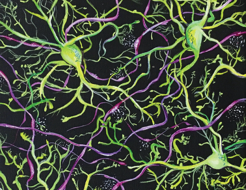
Historically, neuroscience focused on neurons, the functional cellular units of communication in the brain. However, exciting recent advances in microscopy have revealed the importance of many other cell types in essential brain functions. Amongst these supporting players in the brain are microglia, the immune cells of the brain.
Initially, neuroscientists thought that microglia, as immune cells, only act in cases of injury, disease, or infection. However, we now know that microglia play many important roles in brain development and plasticity over the lifespan. Plasticity is the process by which the brain incorporates new experiences into existing neuron connections, in part through physical changes in the connections between neurons. Recently, microglial roles in plasticity have become a focus of research which revealed that microglia interact with neurons to facilitate these physical changes. Following up on this previous work, our study indicated that awake brains inhibit microglial roles in response to brain injury and plasticity.
Neuroscientists long assumed that microglia remain static in the absence of injury or infection. However, the new techniques that allowed visualization of these cells in the living brain revealed that microglia are instead remarkably dynamic. The many branches protruding from each microglial cell body move rapidly through the local brain environment, constantly reaching out and interacting with different components of their surroundings and mediating plasticity. The discovery of microglial dynamics and their participation in plasticity were exciting and raised many questions about how microglia function in the normal healthy brain. Prior to our study, researchers imaged microglia mostly in animals that were anesthetized, meaning a sleep-like state. Instead, we wanted to explore microglia in awake animals and determine whether differences in microglial behavior could influence plasticity.
To study microglia in awake mice, we used a specific type of microscopy which allows us to visualize microglia inside living tissues. Using this technique we, and others in the field of neuroscience, can monitor cell behavior over extended periods. In our study, we imaged the same mice with awake and sleep-like brain states which enabled us to compare how the same brains behave in these two states.
In the sleep-like state, we found that microglial branches spread out and allow microglia to occupy and interact with more of the brain environment whereas such microglial dynamics are greatly reduced in awake brains. We were able to determine the increased concentrations of norepinephrine (a type of adrenaline which transmits signals in the brain) in the awake brain cause microglia to retract their branches and survey less of the brain environment. Importantly, we found that microglia are slower in responding to a brain injury when animals are awake. Additionally, simulating a continuously awake state inhibited plasticity in mice.
Our findings indicate that, in sleep-like states, microglia can better monitor their environment, respond to injuries, and participate in plasticity. These discoveries suggest that if microglia receive prolonged awake signals, they will be less able to help the brain respond to environmental signals that drive both normal brain function and pathological changes. Our study is the first to show that microglia respond to changes in brain state and norepinephrine is the key signal to microglia that the brain is awake.
We concluded that the brain state guides how microglia behave and interact with neurons These findings highlight the importance of healthy sleep-wake cycles as extended wakefulness can prevent normal microglial interactions with neurons and responses to injury. Recognizing how brain state influences microglia will help research on diseases related to altered sleep and awake states, which include neurodevelopmental, neuropsychiatric, and neurodegenerative disorders. Future work can investigate how sleep (rather than anesthetic/sleep-like state) impacts microglia in the brain and what other signals may ultimately regulate their roles in brain health and disease.
Original Article:
Stowell R, Sipe G, Dawes R et al. Noradrenergic signaling in the wakeful state inhibits microglial surveillance and synaptic plasticity in the mouse visual cortex. Nat Neurosci. 2019;22(11):1782-1792.Next read: Stop all the clocks: the hidden long-term consequences of sleep loss by Charlotte N. Hor , Paul Franken
Edited by:
Isa Ozdemir , Senior Scientific Editor
We thought you might like
"Peeling back the onion": a multi-layered approach to understand the dynamics of sleep
Apr 17, 2019 in Neurobiology | 4 min read by Maxime Jan , Paul FrankenVisualizing the effects of sleep on neurons’ maintenance
Oct 11, 2019 in Neurobiology | 3 min read by David Zada , Lior AppelbaumSleep or die: how good sleep decreases the risk of Alzheimer’s disease
Oct 23, 2019 in Health & Physiology | 3.5 min read by Akira OhkuboWhen the girdle of social timing relaxes: Effects of the COVID-19 lockdown on human sleep
Jul 9, 2020 in Psychology | 3.5 min read by Christine Blume , Marlene H. SchmidtMore from Neurobiology
New, smaller-than-ever devices to help us understand how our brain works from the inside
Nov 8, 2024 in Neurobiology | 4 min read by Filippo DonatiCan we use a magnet to see brain inflammation?
Sep 25, 2023 in Neurobiology | 4 min read by Raquel Garcia-Hernandez , Santiago Canals , Silvia de SantisSurprising Behavior Changes in Genetically Modified Syrian Hamsters
Aug 30, 2023 in Neurobiology | 4 min read by Susan Lee , Kim Huhman , Jack TaylorTo achieve goals, we definitively need our neurons
Mar 10, 2023 in Neurobiology | 3.5 min read by Julien CourtinThe Impact of SARS-CoV-2 on the Brain: It Is All in Your Head
Feb 15, 2023 in Neurobiology | 3.5 min read by Meredith G. Mayer , Tracy FischerEditor's picks
Trending now
Popular topics