 Evolution & Behaviour
The Mystery of the Lizard Tail
Quick breakage of lizard tails to escape predation has remained a mystery for centuries. Our study explains the balance between firm attachment and the quick release of the tail and highlights the lizard's way of achieving the “just right” connection for its best chance of survival.

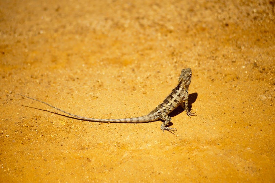
When lizards are under attack, they break off their tails in a fraction of a second and escape. Scientifically, this is known as tail or caudal autotomy. Since the tail is an important organ of lizards for survival as it helps them nourish, run, leap, mate, and escape the next predator, losing the tail is a costly sacrifice. Therefore, in normal circumstances, lizards retain their tails sturdily fixed to the body. But, in life-threatening situations, they can shed them off quickly. This seemingly paradoxical nature of tail attachment has fascinated scientists and remained a mystery for a long time. To answer the question of how the lizard effectively balances its tail's sturdy attachment and quick detachment, we studied the tails of three common lizards, Hemidactylus flaviviridis, Cyrtopodion scabrum, and Acanthodactylus schmidti. Using high-speed videography, we found out that the lizards bent their tails to start the autotomy process along with one of several breaking points, which allowed a specific segment of the tail to be sacrificed rather than losing the whole tail in one go. Surprisingly, the tails did not break when we pulled them straight. It was the bending that triggered the tail's fracture initiation process and ultimately led to a tail loss.
To investigate the tail connection with the body more closely, we imaged the fractured surfaces from both the tail and the body counterpart. On the wedge-shaped "plugs" (found on the tail), we found tightly packed micropillars with mushroom-shaped tops as the ends of muscle fibers. The surface of the mushroom tops had many nanopores. The complementary "sockets" (found on the body) showed the corresponding surface imprints of the mushroom-shaped tops. These surface imprints implied that the mushroom-shaped tops did not penetrate into the surface of "sockets," which would have resulted in a stronger connection. Instead, the lizard has adopted a different strategy for tail attachment with many micro- and nanoscale structural features that created a surface contact with an "intermediate optimal" adhesion strength that was neither too weak nor too strong.
To explore the role of the microscale and nanoscale structural features, we built a biomimetic model. It consisted of an array of micropillars with nanoporous tops using a soft tissue-like elastomer known as polydimethylsiloxane (PDMS). We performed pulling and peeling tests with this model and validated the experimental findings with computational modeling. The main outcomes of our experimental and computational study suggested that the presence of the reported features made the tail-body connections strong via micro- and nanoscale toughening mechanisms, which allowed the tail to attach to the body firmly, avoiding any accidental loss. These toughening features, however, turned out to be highly vulnerable to peeling. The breaking process eased off by 17 times under bending than in non-bending cases. In agreement with the high-speed analysis, bending was clearly the key to quick tail release. Furthermore, the computer model suggested that momentarily contracting muscle fibers around the tail-body connections could accelerate the shedding process along with the bending motion.
Our findings can help bioengineers and scientists to rationally design and optimize tissue adhesives in biomedical areas. Potential applications include adhesive patches that stick to the wet environment inside the body firmly to provide medicinal or therapeutics but come off quickly after treatment. We hope that our biomimetic study encourages other scientists and researchers worldwide to look closely at nature and find more innovative bioinspired solutions to pressing problems in the world.
Original Article:
Baban, N. S., Orozaliev, A., Kirchhof, S., Stubbs, C. J., & Song, Y.-A. (2022). Biomimetic fracture model of lizard tail autotomy. Science, 375(6582), 770–774. https://doi.org/10.1126/science.abh1614Next read: Shrimp on cocaine – what’s the big deal? by Thomas Miller
Edited by:
Isa Ozdemir , Senior Scientific Editor
We thought you might like
From a fossil to a robot…and all the steps in between
Aug 27, 2019 in Evolution & Behaviour | 4 min read by Kamilo Melo , John A. NyakaturaTake Them Outside: Cold Air Helps Croup Symptoms in Kids
Jan 3, 2024 in Health & Physiology | 3.5 min read by Zoé ValbretThe lifetime of memories
Jun 22, 2016 in Neurobiology | 3.5 min read by Thomas Stefanelli , Pablo MendezA message in a bottle dating 250 million years ago
Aug 20, 2019 in Evolution & Behaviour | 3.5 min read by Patrick BlomenkemperMore from Evolution & Behaviour
Cicada emergence alters forest food webs
Jan 31, 2025 in Evolution & Behaviour | 3.5 min read by Martha Weiss , John LillSize does not matter: direct estimations of mutation rates in baleen whales
Jan 29, 2025 in Evolution & Behaviour | 4 min read by Marcos Suárez-MenéndezThe Claws and the Spear: New Evidence of Neanderthal-Cave Lion Interactions
Jan 22, 2025 in Evolution & Behaviour | 3.5 min read by Gabriele RussoA deep-sea spa: the key to the pearl octopus’ success
Jan 20, 2025 in Evolution & Behaviour | 3.5 min read by Jim BarryFeisty fish and birds with attitude: Why does evolution not lead to identical individuals?
Aug 31, 2024 in Evolution & Behaviour | 3 min read by Lukas Eigentler , Klaus Reinhold , David KikuchiEditor's picks
Trending now
Popular topics


